Facilities
Flow cytometry
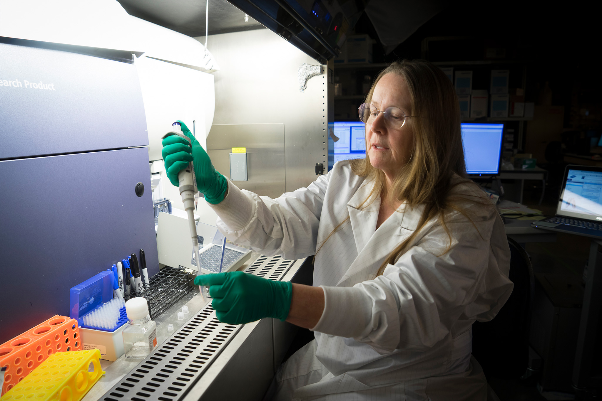
The Flow Cytometry Core offers advanced cell sorting and analysis, including multi-color detection, immunophenotyping, and nanoparticle studies, supported by cutting-edge instrumentation, data analysis tools, and expert staff guidance.
About
Our core facility provides advanced cell sorting and cellular analysis techniques including immunofluorescence detection of 18 colors, cell-cycle distribution, apoptosis measurements, immunophenotyping, plant genotyping, nanoparticle detection, and much more. In addition to state-of-the-art instrumentation and data analysis software, the flow core’s most important resource is the expertise and helpfulness of the staff.
We will help you with experimental design, planning, and execution, as well as subsequent data analysis and the preparation of grant proposals and manuscripts for publication.
Capabilities
Advanced flow cytometry and cell sorting
The core provides high-speed, temperature controlled sorting of sterile, live, and rare cell populations as well as flow cytometry analysis for a variety of applications, including:
- DNA cell cycle analysis
- Apoptosis assays
- Immunophenotyping
- Intracellular phosphoprotein analysis
- Antibody binding evaluation
- Drug Response assays
User training
- We provide training and assistance on how to best design and perform flow cytometry experiments, as well as how to analyze and prepare the resulting data for publication.
- Instrument training on Fortessa and Attune Flow Cytometers
Data Analysis
- FCS Express, FlowJo, and ModFitLt software packages
- Data archiving and offline retrieval
Expert consultation
The core provides a variety of consulting services, including assistance with assay and protocol development, as well as input for grant proposals and manuscripts
Instrumentation
The BD FACS Aria Fusion is a 5-laser, 14-color, 16-parameter high-speed cell sorter with temperature control and housed in an integrated biosafety cabinet. Wavelength excitation choices include 405-nm, 488-nm, 561-nm, 633-nm and 355-nm (UV) lasers. With the advanced gel-coupled cuvette and octagon- and trigon-shaped detector arrays that maximize signal detection, the BD FACSAria III represents the state of the art in cell sorting. In addition, we will also do analysis-only experiments on the FACS Aria that require greater than four colors.
The BD FACS Aria III Cell Sorter is a 5-laser, 14-color, 16-parameter high-speed cell sorter with temperature control and housed within a biosafety cabinet for sorting of biohazardous samples. Wavelength excitation choices now include 405-nm, 488-nm, 561-nm, 633-nm and 355-nm (UV) lasers. Gel-coupled cuvette and octagon- and trigon-shaped detector arrays maximize signal detection. We will do analysis-only experiments on this instrument that require greater then four colors.
The BD Fortessa LSR flow cytometry cell analyzer has the same 5-laser setup as the ARIA cell sorters, with 18-color, 20-parameter detection and a high throughput microwell sample uptake system. This cytometer is available for operation by students and staff. This instrument is used for immunophenotyping, phosphoprotein analysis, DNA analysis, cell cycle and functional assays.
This benchtop flow cytometer has 4-laser excitation, 14-parameter detection capabilities, automated multiwell sampling and automated report generation. Wavelength excitation choices include 405-nm, 488-nm, 561-nm, and 627-nm. This cytometer is available for operation by students and staff. This instrument is used for nanovesicle detection, immunophenotyping, phosphoprotein analysis, DNA analysis, cell cycle and functional assays.
The Chromium system is designed to isolate, process and prepare individual cells for genome and transcriptome analysis. The instrument is used to sort cells in liquid suspension into 96 separate chambers to prepare them for DNA or RNA extraction. Researchers apply NGS to identify changes in the genome or the gene expression patterns that affect cellular events and metabolic processes.
The facility includes an upright Nikon Optiphot fluorescence microscope and an inverted Olympus microscope. Two well-equipped BSL-2 tissue culture facilities adjoin the flow core, for sterile processing of live cells, both pre- and post-sort. The flow cytometry data analysis support includes software licenses for FCS Express and FlowJo for general listmode data analysis. These data analysis programs are available for use without charge on two workstations within the laboratory. Flow core staff are available to help users with experimental design, sample preparation, sample running, cell sorting, and data analysis. Trained users have 24-hour open access to the facility.
Rates
| Service | Internal | PCCR Subsidized | External Non-Profit and Small Business | External for Profit |
|---|---|---|---|---|
| FACS Aria Cell Sorters | $47/hr | $29/hr | $194/hr | $207/hr |
| Fortessa Cell Analyzer | $43/hr | $32/hr | $132/hr | $140/hr |
| Attune Nxt Cell Analyzer | $43/hr | $32/hr | $132/hr | $140/hr |
| 10x Chromium | $450/run | $350/run | $1084/run | $1154/run |
| Flow Cytometry Staff Time | $70/hr | $50/hr | $151/hr | $161/hr |
| Training | Instrument time charge | Instrument time charge | Instrument time charge | Instrument time charge |
| Consultation | Free | Free | Free | Free |
Resources
Developed by Justin Meyers
Sample: Cell line or primary cells
Materials
- Propidium iodide reagent (Sigma P-4170) 10mg make up 1mg/ml solution in DI water and store at 4C
- Rnase A, (Sigma R-6513), 10mg, store at -20C
Procedure
Isolate cells
- Harvest cells, want good single cell suspension
- Count cells
- Dispense aliquots of 1 million cells into labeled tubes
- Add 5 ml PBS and centrifuge for 5 min at 400 x g.
- Re-suspend cell pellet in 300 ul PBS with gentle vortex
Fix cells
- Add 700 ul ice cold ethanol (200 proof), slowly, while continuously vortexing.
- Incubate at 4C for 30 min to overnight
- Pellet cells at 2000 rpm for 5 min
- Re-suspend (vortex gently) cells in 250 ul of PBS
- Add 5ul of 10mg/ml Rnase A (final concentration being 0.2-0.5 mg/ml)
- Incubate at 37C for one hour
- Add 10ul of the 1mg/ml PI solution (final concentration is 10ug/ml)
- Keep in the dark for at least one hour until analysis
- Perform flow cytometry analysis on flow
Developed by Justin Meyers
Sample: Cell line or primary cells
Materials
- Directly conjugated antibodies
- FACS buffer (DPBS + 5% FBS)
- BD Cytofix (BD 554655) or 1% paraformaldehyde
Procedure
Isolate cells
- Harvest cells, want good single cell suspension
- Count cells
- Dispense aliquots of 1 million cells into labeled tubes
- Add 5 ml FACS Buffer and centrifuge for 5 min at 1300 RPM
- Re-suspend cell pellet in 100ul FACS Buffer with gentle vortex
Fix cells
- Add appropriate amount of antibody
- Vortex gently
- Incubate at 4C for 30 min
- Add 4 ml of FACS Buffer
- Pellet cells at 1300 rpm for 5 min
- Re-suspend cells in 500 ul of FACS Buffer
- Keep cells on ice up to 1 hour before analysis*
- Analyze cells on flow cytometer
*If cells cannot be analyzed within one hour, fix the cells in 0.5 ml of 1X BD Cytofix or 1% paraformaldehyde and store at 4C for analysis the following day
Developed by Justin Meyers
Sample: Cell line or primary cells
Materials
- Cells stained with antibody
- BD Cytofix (BD 554655)
- D-PBS
- FBS
Procedure
Procedure for fixing cells with BD Cytofix™
- Pellet 106 cells by centrifugation (250 – 300 x g) and carefully remove supernatant
- Make up 1X of fixation buffer by adding 5 ml of Cytofix (BD554655) to 10 ml of DPBS
- Add either 300μl (for microwell plates) or 500 μl (for tubes) aliquots of 1X fixation buffer to each cell pellet and resuspend the cells by either pipetting or vortexing.
- Incubate the cells with fixation buffer for 15 to 30 min at 4°C. (Cell aggregation can be avoided by vortexing prior to the addition of the fixation buffer.)
- Fixed cells should be washed and suspended in a buffer that contains protein. (DPBS + 5% FBS) for longer term storage. They can be left in the fixative for up to two days.
Store the fixed cells at 4°C (protected from light).
It is recommended that fixed cell samples be read as soon as possible, i.e., within one week.