Facilities
Light and fluorescence microscopy
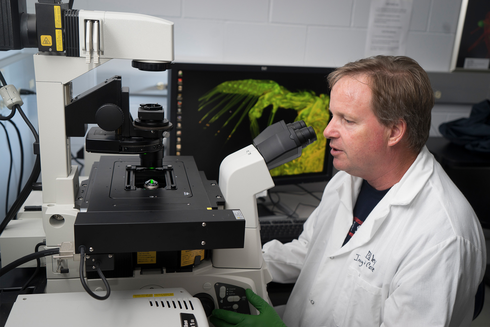
Purdue’s Imaging Facility provides advanced light microscopy, including confocal, multi-photon, and light-sheet imaging, plus pre-clinical bioluminescence, fluorescence, and CT, supporting research and development for Purdue and the broader academic and non-academic community.
About
Purdue’s imaging facility is a research resource for Purdue faculty, staff, students, and other academics and non-academics seeking access to state-of-the-art light microscopy for their research and development work. We specialize in confocal, multi-photon, and light-sheet microscopy, as well as pre-clinical bioluminescence/fluorescence and computed tomography.
We serve researchers at Purdue University and biotech companies in the community as a member of the Indiana CTSI service core community. We provide the instruments and expertise needed to visualize molecules in preparations ranging from single cells to entire animals. All facility users receive individualized instrument training and project-specific advice for optimal data acquisition. Consultation on sample preparation, image rendering, and data analysis is also available as our knowledge base permits. We encourage collaborations and contract research services.
capabilities
Confocal microscopy
Our facility provides three visible-wavelength laser point scanning confocal systems suitable for creating multi-dimensional images of living or non-living samples.
- Multi-colored fluorescence imaging
- Photoablation/photoactivation
- Imaging in aqueous environments
- Material science – fluorescence and reflectance
Multiphoton microscopy
If your sample is thicker than 100 µm or you require less phototoxicity to your living sample, you might benefit by using a multi-photon (MP)
- Multi-dimensional images of thick samples, up to 800 microns
- Live cell and live animal (intravital) imaging
- Photo-uncaging
- Photoablation/photoactivation
- Second Harmonic Generation (non-centrosomatic)
TIRF microscopy
Total Internal Reflection Fluorescence uses a finite wave-front of energy to fluorescently illuminate samples and limit excitation of fluorophores close to the coverslip to dramatically increase the signal to noise of your sample.
- Protein dynamic studies on the cell membrane
- Single molecule examinations
Super Resolution microscopy
For spatial resolution beyond the diffraction-limit (<220 nm) we offer Super Resolution microscopy with both SIM (3 colors) and STORM.
- Very high resolution intracellular imaging in thin samples
- Detailed localization analysis
Wide-field imaging
- Very high resolution intracellular imaging in thin samples
- Detailed localization analysis
Pre-clinical molecular and microCT
The Bindley Imaging Facility provides systems capable of imaging larger subjects such as small rodents, medical devices or other non-biologics
- Small Animal CT Imaging
- Material science – Computed Tomography
- Fluorescent and Bioluminescent Imaging
Image processing and analysis for all data
Images are great to display but have so much more data. Use one of our commercial analysis software tools to pull out all the information.
- Co-localization analysis
- Intensity analysis
- Time-lapse imaging of dynamic intracellular events
- Three-dimensional morphology
- Deconvolution
Our team is dedicated to helping facility users maximize the value of their imaging data. We offer:
- Training on software tools and analysis workflows
- Consultation for experimental design and data interpretation
- Custom pipeline development and scripting support for specialized or high-throughput analysis needs
Light Sheet Microscopy
Light sheet microscopy excels at capturing fast 3D imaging of living or optically cleared samples with minimal light exposure:
- Rapid volumetric imaging with minimal phototoxicity and bleaching
- Multi-view acquisition for isotropic resolution
- Compatible with live embryos, organoids, spheroids, small organisms, and cleared tissue samples
- High temporal resolution for developmental and dynamic processes
- Long-term imaging of living samples
Instrumentation: Microscopy
- Inverted microscope (Nikon TiE based)
- Motorized XY stage
- Piezo z-drive and standard TiE z-motor
- Laser: 445 nm, Argon: (457 nm, 476 nm, 488 nm, 514 nm), 561 nm, 640 nm
- Four different PMT detectors or 32-channel spectral detector
- Transmitted detector for DIC-like confocal imaging
- Reflectance imaging
- Hybrid scanner (galvanometer and resonant – up to 30 fps)
- Live imaging with Tokai Hit stage-top chamber
- Perfect focus system keep area of interest in focus
- NIS-Elements software encompasses acquisition through analysis
- FRAP and FRET experiment possible
- Nikon Objective Choices
For additional detailed specifications, please contact the facility manager.
Location: Bindley Bioscience Center
- Inverted micrscope (Nikon TiE based)
- Motorized XY stage
- Standard TiE Z-motor
- 405 nm, Argon: (457 nm, 476 nm, 488 nm, 514 nm), 561 nm, 640 nm
- Tunable Spectra Physics Mai Tai DeepSee Ti:Sapphire laser (690-1040nm) for Multi-Photon applications and stimulation
- Four different PMT detectors
- Transmitted detector for DIC-like confocal imaging
- Hybrid scanner (galvanometer and resonant – up to 30 fps)
- Live imaging with Tokai Hit stage-top chamber (inquire about dish compatibility)
- NIS-Elements software encompasses acquisition through analysis
- Nikon Objective Choices
For additional detailed specifications, please contact the facility manager.
Location: Bindley Bioscience Center
- Upright microscope (Nikon FN-1)
- Dual Coherent IR lasers (tuneable 800 – 1300 nm and fixed 1040 nm) for multi-photon imaging
- Three GaAsP and one high-sensitive red non-descanned detectors
- Second Harmonic Generation for imaging collagen
- Anesthesia equipment and heat plate for animal imaging
- Visible lasers 405 nm, 488 nm, 561 nm, 640 nm for standard or optogenetics imaging
- Spectral GaAsP Detector for standard visible confocal
- Hybrid scanner (galvanometer and resonant – up to 30 fps)
- Motorized Prior XYZ
- High-speed piezo-Z motor
- Prior Scientific Z-Deck motorized stage
- Nikon Objective Choices
For additional detailed specifications, please contact the facility manager.
Location: Bindley Bioscience Center
- Inverted microscope (Nikon TiE based)
- Motorized stage
- Piezo z-drive
- Perfect focus system
- STORM (or cSTORM) with 647nm, 561nm and 488nm lasers
- SIM with 405nm, 488nm and 561nm lasers
- TIRF capability (multicolor TIRF (10 ms/channel))
- Nikon Objective Choices
For additional detailed specifications, please contact the facility manager.
Location: Bindley Bioscience Center
- Upright microscope (Nikon 90i based)
- Differential Interference Contrast (DIC) and standard polarizers
- Brightfield
- Nikon DS-Ri1 12MP color camera
- Manual stitching with NIS-Elements Software
- Nikon Objective Choices
For additional detailed specifications, please contact the facility manager.
Location: Bindley Bioscience Center
- Inverted microscope (Nikon TiS)
- Phase contrast and brightfield
- Zenon light source for fluorescence imaging
- High sensitive Photometrics QuantEM EMCCD camera
- Standard fluorescent filter cubes including unique far-red filters
| Far-Red Filter # | Excitation [nm] | Emission [nm] |
|---|---|---|
| 1 | 590-650 | 663-738 |
| 2 | 725-775 | 780 LP |
| 3 | 750-800 | 810-860 |
For additional detailed specifications, please contact the facility manager.
Location: Bindley Bioscience Center Room 233
- Upright microscope (Zeiss Axio Examiner Z1)
- Laser: 405 nm, Argon: (458 nm, 488 nm, 514 nm), 561 nm and 633 nm.
- Detectors:
- 32-channel GaAsP spectral photomultiplier tube (PMT)
- 2 MA-PMT for blue and red channels
- Transmitted light PMT
- Unique objective turret for both standard and dipping applications
| Objective | Magnification | NA | Cover Glass thickness | Working distance (mm) | Media | Condensor Prism |
|---|---|---|---|---|---|---|
| Plan Apochro mat | 5x | 0.16 | 0.17 | 1231 | dry | NA |
| Plan Apochro mat | 10x | 0.45 | 0.17 | 2 | dry | II |
| W-Plan Apochro mat | 20x | 1 | none | 2.4 | water dipping | III |
| Plan Apochro mat | 20x | 0.8 | 0.17 | 0.55 | dry | II |
| W-Plan Apochro mat | 20x | 1 | 0.17 | 1.7 | water immersion | III |
| LD C-Apochro mat | 40x | 1.1 | 0.17-0.19 | 0.62 (at 0.17) | water immersion | III |
| W-Plan Apochro mat | 40x | 1 | none | 2.5 | water dipping | III |
| Plan Apochro mat | 63x | 1.4 | 0.17 | 0.19 | oil | III |
For additional detailed specifications, please contact the facility manager.
Location: Hansen Lifescience Building
The lightsheet system is perfect for capturing 3D fluorescent images of organisms, tissue explants, and cell cultures over long periods. Basically, it shines a thin sheet of laser light on a slice of your sample, and high-speed cameras take pictures of the glowing parts from the side. Using the light sheet method means less damage to your sample from laser exposure, which is great for imaging delicate things like embryos, larvae, and organoids. You can use it to image alive, preserved, or optically cleared samples.
- Two Nikon CFI Plan Fluor 10x W 0.3 NA water immersion objective lenses for illumination.
- Two Olympus XLUMPLFLN 20x (eff. 22.2x) W 1.0 NA water immersion objective lenses for detection
- Two high-speed Hamamatsu Orca Flash 4.0 V3 sCMOS cameras
- Laser wavelength: 405 nm, 488 nm, 515 nm, 561 nm, 642 nm, 785 nm
- Flexible light-sheet thickness (2 µm to 4 µm)
- Filters:
- Bandpass 418-462, 457-501, 500-530, 526-564, 580-627, 655-704
- Longpass 498
- Photomanipulation module: 532 nm ablation
For additional detailed specifications and sample preparation, please contact the facility manager.
Location: Hansen Lifescience Building B032
Instrumentation: Pre-clinical
The Ami can perform either Fluorescence or Bioluminescent Imaging with a standard photography overlay.
- 25 x 17 cm to 10 x 7 cm Field of View
- Imaging Platform 56 x 66 x 122 cm3 (width x depth x height)
- Heated imaging platform
- Free vendor software available for download
| Exitation [nm]: | 430 | 465 | 500 | 535 | 570 | 605 | 640 | 675 | 710 | 745 |
| Emission [nm]: | 530 | 650 | 670 | 690 | 710 | 750 | 770 | 790 | 810 | 830 |
(All values ± 10 nm range)
For additional detailed specifications, please contact the facility manager.
Location: Hansen Lifescience Building
- 72 mm Field of View
- Down to 4.5 µm resolution after sub or slice reconstruction
- Max. tube voltage 90 kV
- Max. tube current 200 µA
- Max. output 8 W
- 60 fps
- Invivo imaging capability for different animals
For additional detailed specifications, please contact the facility manager.
Location: Hansen Lifescience Building
Nuclear medicine pre-clinical instrument for PET/SPECT/CT.
- Resolution 0.35 – 0.8 mm based upon collimator
- Multi-isotope imaging (20 keV – 511 keV)
- Auto-fusion of CT images
- Interchangeable and easy-to-mount collimators
For additional detailed specifications, please contact the facility manager.
Location: Hansen Lifescience Building
Instrumentation: Image Analysis
All workstation computers can be accessed either in person or remotely through TeamViewer. They are free to use, but advance reservation in iLab is required to use the software.
Rates
| Service | Internal Customers | PCPB Customers | External Non-Profit & Small Business | External For Profit |
|---|---|---|---|---|
| Nikon A1RMP-HD Intravital Confocal | $54/hr | $54/hr | $108/hr | $115/hr |
| Perkin Elmer Micro-CT | $48/hr | $48/hr | $181/hr | $192/hr |
| Spectral Ami Optical Imaging System | $39/hr | $39/hr | $68/hr | $73/hr |
| MiLabs U-SPECT-II/CT/PET | $72/hr | $72/hr | $163/hr | $173/hr |
| Nikon A1R-MP | $53/hr | $37/hr | $83/hr | $88/hr |
| Nikon A1Rsi | $40/hr | $28/hr | $132/hr | $141/hr |
| Super Resolution Nikon N-STORM/N-SIM/TIRF | $52/hr | $37/hr | $82/hr | $87/hr |
| Zeiss LSM 880 | $38/hr | $29/hr | $71/hr | $76/hr |
| Zeiss LSM 880 – after hours | $28/hr | $26/hr | $44/hr | $47/hr |
| Nikon 90i Wide-Field | $0/hr | $0/hr | $0/hr | $0/hr |
| Nikon TiS Wide-Field | $0/hr | $0/hr | $0/hr | $0/hr |
| Training: Confocals, Super Resolution, VECTor/CT | $224 | $152 | $347 | $347 |
| Training: AMI, Widefield, Plate Readers | $124 | $124 | $192 | $192 |
| Synergy Neo Plate Reader | $15/hr | $15/hr | $23/hr | $24/hr |
| DynaPro II Plate Reader | $10/hr | $10/hr | $17/hr | $18/hr |